

IB Syllabus Requirements for Cell and nuclear division
D2.1.1
Generation of new cells in living organisms by cell division
D2.1.2
Cytokinesis as splitting of cytoplasm in a parent cell between daughter cells
D2.1.3
Equal and unequal cytokinesis
D2.1.4
Roles of mitosis and meiosis in eukaryotes
D2.1.1
New cells come from existing cells
Cell division is a biological process in which one parent cell divides to form daughter cells. In all living organisms, new cells are made by division of a pre-existing cell; in normal life, we don’t see fully functioning cells being built from non-living chemicals.
A parent cell is a cell that divides to produce new cells. A daughter cell is a cell formed by division of a parent cell. You may also see the term “mother cell”, but the wording can be a bit misleading: the parent cell does not stay behind as a separate individual after division. It becomes the two daughter cells.
Cell division does three major jobs: growth, maintenance and reproduction. A multicellular organism grows when its cell number increases. It maintains tissues by replacing cells that are lost or damaged, and it may reproduce by producing cells that form offspring. In asexual reproduction, repeated mitotic divisions can produce large numbers of genetically identical cells, so a clone can behave as a single genetic individual even when it looks like many separate parts.
D2.1.2
Dividing the cytoplasm
Cytokinesis is the division of the cytoplasm of a parent cell between daughter cells. It differs from nuclear division: mitosis or meiosis divides nuclei; cytokinesis divides the cell body.
In animal cells, cytokinesis depends on a contractile ring just inside the plasma membrane. Actin and myosin proteins make up the ring. As it contracts, the ring pulls the membrane inward at the cell equator and forms a cleavage furrow. Once the furrow reaches the centre, the cell is pinched into two.
Plant cells can’t simply pinch inwards because a rigid cell wall gets in the way. Vesicles collect at the equator of the cell, then fuse to form new plasma membrane between the daughter cells. Materials carried in vesicles also help build the new cell wall between them. The key comparison is simple: animals pinch the membrane inwards; plants build a separating plate outwards across the middle.

D2.1.3
Equal division is common, but not universal
Equal cytokinesis is cytokinesis where the cytoplasm is shared in roughly equal amounts between daughter cells. It’s common in tissues where the two daughter cells will do similar jobs, for example in a growing root tip.
Unequal cytokinesis is cytokinesis where one daughter cell gets much more cytoplasm than the other. The smaller daughter cell can survive only if it gets a nucleus and at least one of each organelle that cannot be made from scratch. Mitochondria are the classic example: cells make new mitochondria by the growth and division of mitochondria that already exist, so a daughter cell with none would be in serious trouble. The same idea applies to chloroplasts in plant cells that need them.
Budding in yeast
Budding is a form of asexual reproduction where a smaller daughter cell grows out from a larger parent cell. In yeast, the nucleus divides by mitosis, one nucleus moves into the bud, and a wall forms between the bud and the larger cell. Because the bud receives only a small share of the cytoplasm, this counts as unequal cytokinesis.
Oogenesis in humans
Oogenesis is the production of egg cells in ovaries by meiotic divisions and cell differentiation. In human oogenesis, cytokinesis is strongly unequal. Most of the cytoplasm stays in one large cell that can become an oocyte. The tiny polar bodies receive nuclei but very little cytoplasm, and usually do not develop further. This fits the biology: the egg cell needs reserves for early development, while the polar bodies discard extra chromosome sets.

D2.1.4
Nuclear division must come before cell division
Nuclear division is the division of a nucleus, with genetic material shared out into new nuclei. If a eukaryotic cell split its cytoplasm before dividing its nucleus, one daughter cell could end up with no nucleus at all. An anucleate cell is a cell that lacks a nucleus. Without a nucleus, the cell cannot transcribe nuclear genes, so it cannot make the full range of proteins needed for long-term growth and maintenance.
Mitosis is nuclear division that produces genetically identical nuclei while maintaining chromosome number. Eukaryotes use it for growth, replacement of cells, tissue repair and asexual reproduction. Mitosis preserves the genome: each daughter nucleus receives the same genes as the parent nucleus.
The symbol is the haploid chromosome number, where is the number of different chromosome types in one complete set (dimensionless count). A diploid eukaryotic body cell has chromosomes. When a diploid nucleus undergoes mitosis, it produces diploid daughter nuclei, so the chromosome number is maintained.
Meiosis is nuclear division that halves chromosome number and generates genetically varied nuclei. It is used in sexual life cycles to produce nuclei that can contribute to gametes. Meiosis changes a diploid nucleus into haploid nuclei, so fertilization can restore the diploid number instead of doubling the chromosome number every generation.

Organism growth depends on several processes working together. Cells divide by mitosis, cytokinesis splits the cytoplasm, and the resulting cells grow and specialize. In plants, enlargement of cells and production of biomass also matter, but mitosis and cytokinesis supply the new cells on which those processes act.
D2.1.5
Chromosomes must be copied first
DNA replication is the synthesis of a new DNA molecule using an existing DNA molecule as a template. Before mitosis or meiosis begins, the cell replicates all nuclear DNA, so genetic information can be passed on to daughter nuclei.
A chromosome is a DNA molecule associated with proteins that carries a linear sequence of genes. Once DNA replication has happened, each chromosome contains two DNA molecules. A chromatid is one of the two DNA molecules of a replicated chromosome. Sister chromatids are chromatids produced by replication of the same chromosome, so they are genetically identical unless a mutation has occurred.
The cell keeps sister chromatids together until anaphase. A cohesin is a protein complex that holds sister chromatids together after DNA replication. When cohesin is cut at the correct time, the chromatids can separate and move to opposite poles.

Here’s a useful exam phrase: after replication, a chromosome has two chromatids, but it is still counted as one chromosome until the chromatids separate. Once they separate, each chromatid is considered a chromosome in its own right.
D2.1.6
Making DNA manageable
Chromosome condensation is the packaging of long DNA molecules into shorter, thicker chromosome structures for nuclear division. It’s needed because DNA molecules are extremely long compared with the nucleus. Left fully extended, they would tangle, snap or fail to separate cleanly.
Histones are proteins that DNA winds around to form nucleosomes. This wrapping compacts DNA. Extra coiling and folding shorten it further, producing visible chromosomes during nuclear division.
Moving chromosomes
Microtubules are hollow protein fibres made from tubulin that can rapidly assemble and disassemble. During mitosis and meiosis, they form a spindle that extends from the poles of the cell towards chromosomes.
A kinetochore is a protein structure on a chromosome where spindle microtubules attach. It also works as a microtubule motor: by removing tubulin subunits, it helps shorten attached microtubules and pull chromosomes or chromatids towards poles.
The basic sequence is straightforward. Chromosomes condense first, so they can be moved safely. Then microtubules attach and pull. In mitosis and meiosis II, sister chromatids separate. In meiosis I, homologous chromosomes separate.

D2.1.7
The four phases
Mitosis is usually split into four phases: prophase, metaphase, anaphase and telophase. Interphase is not part of mitosis, although DNA has already been replicated during interphase before mitosis starts.
Prophase
During prophase, chromosomes condense and the mitotic spindle begins to form. By the end of prophase, the nuclear membrane breaks down, which lets spindle microtubules interact with the chromosomes.
Metaphase
In metaphase, chromosomes line up at the equator of the cell. Sister chromatids attach to opposite spindle poles, and tension checks that the attachment is correct. Cohesin still holds the sister chromatids together.
Anaphase
In anaphase, sister chromatids separate and move to opposite poles. Cohesin is cut, and kinetochore microtubules shorten. From this point on, the separated chromatids are chromosomes.
Telophase
In telophase, chromosomes reach the poles, nuclear membranes reform and the chromosomes decondense. Cytokinesis often starts during telophase, so by the end of mitosis the cell is usually already well on its way to becoming two cells.

The process produces two genetically identical daughter nuclei because DNA was copied before mitosis, then one copy of each chromosome moves to each pole.
D2.1.8
What to look for in diagrams and micrographs
When you identify mitotic phases, look first at what the chromosomes are doing. Cell shape by itself can be misleading.
- Interphase: nucleus visible; chromosomes not individually distinct.
- Prophase: chromosomes visible as condensing threads or rods; not aligned on the equator.
- Metaphase: chromosomes aligned across the equator.
- Anaphase: two groups of chromosomes separating towards opposite poles; often V-shaped or pulled-looking.
- Telophase: two chromosome groups at poles; nuclei reforming; cytokinesis may be visible.
Plant root tips are useful for practice because the cells often sit in files. To make a root tip squash, roots are grown, the tissue is fixed, acid is used to loosen the cell walls, DNA is stained so the chromosomes can be seen, and the root tip is gently squashed under a coverslip before viewing under a microscope. Take care with the acid and stains: they require eye and skin protection.
The same skill works for drawn diagrams and for real cells seen through a microscope or in a micrograph. Real cells rarely look as tidy as textbook drawings. Use several clues together: chromosome condensation, alignment, separation and whether one or two nuclei are visible.

D2.1.9
Haploid and diploid
A haploid nucleus contains one complete set of chromosomes, with no homologous pairs. A diploid nucleus contains two complete sets of chromosomes, arranged as homologous pairs.
A homologous chromosome pair is a pair of chromosomes with the same genes in the same sequence, though the alleles may differ. One chromosome in the pair may have come from the maternal parent, and the other from the paternal parent.
A reduction division is a nuclear division that halves the chromosome number. Meiosis counts as a reduction division because one diploid nucleus ultimately gives four haploid nuclei.
Why meiosis is needed
In a sexual life cycle, fertilization fuses two haploid gametes to form a diploid zygote. If gametes were diploid, the chromosome number would double every generation. Meiosis prevents that by producing haploid nuclei before gamete formation or at another stage in the sexual life cycle.
Two rounds of segregation
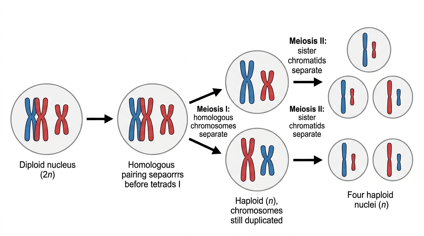
Segregation is the separation of chromosomes or chromatids to opposite poles during nuclear division. Meiosis has two rounds of segregation:
- In meiosis I, homologous chromosomes separate. A diploid nucleus becomes haploid nuclei because each new nucleus receives only one chromosome from each homologous pair.
- In meiosis II, sister chromatids separate. This is similar to mitosis, but it happens in haploid cells and produces four haploid nuclei in total.

D2.1.10
When chromosomes fail to separate
Non-disjunction is an error in nuclear division where homologous chromosomes or sister chromatids do not separate correctly. In meiosis I, a homologous pair may travel to the same pole. In meiosis II, sister chromatids may travel to the same pole instead.
This produces gametes with an abnormal chromosome number: either one chromosome too many or one chromosome missing. A missing chromosome is usually lethal because too many essential genes are absent. An extra chromosome is often lethal too, although some cases survive.
Down syndrome is a human genetic condition usually caused by having three copies of chromosome 21. This is called trisomy 21. It can happen when a gamete carrying an extra chromosome 21 fuses with a normal gamete. Individuals vary, but possible features include effects on learning, hearing, vision and heart development.

Non-disjunction can also affect sex chromosomes, giving combinations such as or a single chromosome. For this topic, don’t try to memorize every condition. Focus on the meiotic error: chromosomes did not segregate correctly.
D2.1.11
Meiosis reshuffles alleles
Meiosis produces genetic diversity by making new combinations of alleles. Two mechanisms matter here: crossing over and random orientation of bivalents.
A bivalent is a pair of homologous chromosomes associated together during meiosis I. Synapsis is the pairing of homologous chromosomes to form bivalents. By this stage each chromosome has already replicated, so a bivalent contains four chromatids.
Crossing over is the exchange of corresponding DNA segments between non-sister chromatids of homologous chromosomes. It happens during prophase I. At the points where chromatids cross and stay attached, a chiasma is visible; a chiasma is a physical connection between homologous chromatids produced by crossing over.
Crossing over makes recombinant chromatids: chromatids with allele combinations not present in either original homologous chromosome. Because crossing over can happen at different positions along chromosomes, it adds a lot of variation.

Random orientation of bivalents is the chance alignment of each homologous pair relative to the poles during metaphase I. For each bivalent, either homologous chromosome can face either pole. One bivalent’s orientation is independent of the orientation of the others.
The number of possible chromosome combinations from random orientation is , where is the haploid chromosome number, already defined as the number of chromosome types in one complete set. In humans, , so random orientation alone can produce over eight million chromosome combinations before crossing over is even considered.

This is one way sexual reproduction supplies variation for evolution. Meiosis creates varied gametes; fertilization combines gametes; natural selection can then act on the resulting variation in phenotypes. Variation does not guarantee evolution in a particular direction, but without heritable variation there is little for selection to sort.
D2.1.12
Increasing cell number
Cell proliferation means an increase in cell number because cells divide faster than they are lost. In multicellular eukaryotes, proliferation relies on repeated mitosis followed by cytokinesis: the genome is copied and shared out before the cells separate.
Growth
In early animal embryos, repeated cell division builds the embryo from a single zygote into many cells. These early divisions are especially noticeable because they increase cell number quickly and produce cells that can later specialize.
In plants, growth is focused in meristems, regions of undifferentiated plant tissue where cells keep dividing. Root and shoot apical meristems make new cells that lengthen roots and shoots. Some daughter cells stay in the meristem and continue dividing; others leave, enlarge and differentiate.

Replacement and repair
Routine cell replacement occurs in tissues that continually lose cells. Skin is a good example: deeper layers produce new cells, while older cells are eventually shed from the surface.
Tissue repair depends on cell division too. During wound healing in skin, surviving cells close to the wound divide and replace lost cells. If the damage is too severe and the relevant dividing cells have been lost, repair becomes much harder.
So, for the broader growth question: organisms grow through coordinated cell proliferation, cell enlargement, biosynthesis of cell materials and differentiation. Cell division provides more cells; growth processes then give those cells their size and function.
D2.1.13
The repeating cycle used for proliferation
The cell cycle is the ordered sequence of events by which a cell grows, replicates its DNA, divides its nucleus and divides its cytoplasm. Cell proliferation happens as cells go through this cycle again and again.
Interphase is the period of the cell cycle between one mitosis and the next. It includes , and .
G1 phase is the stage of interphase after mitosis and before DNA replication, when the cell grows and carries out normal metabolism. Each chromosome consists of one DNA molecule.
S phase is the stage of interphase during which nuclear DNA is replicated. After S phase, each chromosome consists of two sister chromatids held together.
G2 phase is the stage of interphase after DNA replication and before mitosis, when the cell continues preparation for nuclear division.
After G2, the cell enters mitosis, then cytokinesis. Some cells leave the repeating cycle and enter , a non-dividing state in which they may differentiate and carry out a specialized function.

DNA content per nucleus can help identify the cell-cycle stage. Cells in have the lower DNA content, cells in and early mitosis have about double that DNA content, and cells in phase fall between these values because replication is in progress.
D2.1.14
Interphase is busy, not resting
Interphase is metabolically active. The cell may not be visibly dividing, but plenty is happening to prepare it for division later.
During interphase, growth depends on biosynthesis: the enzyme-controlled production of cellular molecules from simpler substances. The cell builds proteins, including enzymes and structural proteins. In S phase, it also makes DNA, while membrane components such as phospholipids and membrane proteins are produced as well.
The cytoplasm increases in volume, and the cell must increase its number of organelles before it divides. Mitochondria and chloroplasts matter especially here, because new ones come from the growth and division of pre-existing mitochondria and chloroplasts. A cell with none cannot just assemble them from loose parts.
Chromatin state matters too. Chromatin is DNA associated with proteins in a less condensed form within the nucleus. When chromatin is decondensed, it can be transcribed, which allows protein synthesis. Some DNA stays more condensed as heterochromatin, where genes are not needed for that cell’s current activities.

D2.1.15
Cyclins rise and fall
Cyclins are regulatory proteins that rise and fall in concentration during the cell cycle, helping the cell move through checkpoints. You don’t need the detailed roles of named cyclins for the syllabus. Keep it at the right level: different cyclins peak at different times.
A checkpoint is a control point in the cell cycle where the cell can progress only when specific conditions have been met. A cell passes a checkpoint when the relevant cyclin reaches a threshold concentration. If that threshold is not reached, the cell does not move on to the next stage.
This control matters because cell division is powerful. Too little proliferation prevents growth and repair; too much proliferation risks tumour formation. Cyclins help keep the sequence orderly: growth, DNA replication, preparation for mitosis, mitosis and cytokinesis.

D2.1.16
When control genes mutate
A mutation is a change in the base sequence of DNA. When mutations affect genes that control the cell cycle, cells may no longer divide at the right time or stop when they should. If one cell loses that control, it can keep dividing, and its daughter cells inherit the same faulty control.
A proto-oncogene is a normal gene that promotes cell division or cell-cycle progression when appropriately regulated. An oncogene is a mutated or overactive form of a proto-oncogene that stimulates excessive cell division. Just one overactive copy may raise the risk, since the faulty signal actively drives the cell cycle forward.
A tumour suppressor gene is a gene whose normal product inhibits cell division, supports DNA repair or promotes death of badly damaged cells. When tumour suppressor genes lose function, the cell’s division “brakes” become weaker. In most cases, both copies have to be affected before control is seriously lost, because one working copy may still make enough functional protein.
Mutagens, including some chemicals and high-energy radiation, increase mutation rates. That raises the chance of damage to cell-cycle control genes. Tumour formation usually needs several mutations to build up in the same cell lineage, rather than one unlucky change. Every division of a partly deregulated cell produces more cells in which further mutations can occur.
D2.1.17
Tumours are not all the same
A tumour is a mass of cells formed by uncontrolled cell division. Tumours can differ quite a lot: some have cells dividing quickly, some grow faster than others, and some stay as one mass while others spread.
A primary tumour is the original tumour at the site where uncontrolled division first began. A benign tumour is a tumour that remains localized and does not invade neighbouring tissues or spread to distant sites. Benign tumours are not classed as cancer, though they can still cause problems if they press on tissues.
A malignant tumour is a tumour whose cells can invade nearby tissues and spread to other parts of the body. Metastasis is the spread of tumour cells from one part of the body to another. A secondary tumour is a tumour that forms after metastatic cells settle and divide at a new site. Malignant tumours are cancers.

Mitotic index
The mitotic index is the ratio of cells in mitosis to the total number of observed cells in a sample. It is calculated as:
To work it out, look at a population of cells in a micrograph or microscope field. Count the total number of cells, count the cells in mitosis, then divide. In practice, this could be done using a root tip meristem or a tumour micrograph. A higher mitotic index shows that a larger fraction of cells are actively dividing at that moment, which often suggests more rapid proliferation.

Cancer treatments such as chemotherapy often target mitosis because cancer cells divide frequently. The snag, and it is a serious one, is that normal rapidly dividing cells can also be damaged, which explains many side effects.
Were those notes helpful?
The Fast Track To Your
Best IB Coursework & College Essays
All content on this website has been developed independently from and is not endorsed by the International Baccalaureate Organization. International Baccalaureate and IB are registered trademarks owned by the International Baccalaureate Organization.